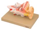
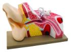
Enlarged approximately 4 times. The petros portion of the temporal bone section of the auditory canal are removable, with labyrinth can be taken out and opened. The tympanic membrane with malleus and incus can be removed in 5 parts.
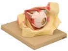
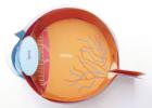
Model shows both sides of an eye, enlarged 5 x. One side of the model shows the eye socket with a sagittal cutaway and the background to the eye and the electron microscopic fine structure of the retina are shown separately.
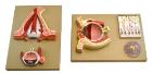
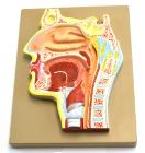
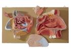
Enlarged 2 times, this cross section of human head to show mouth and nasal passages. Relationship between the windpipe and esophagus can be easily shown. Includes key card
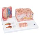
Model Microanatomy* Tongue, specific parts of the tongue, macroscopic view of the tongue in life size (dorsal view) and microscopic views of the various papillae of the tongue (10-20x life size) and of a taste bud ( 450x life size), Dimensions : 14.5 x 32.5 x 20 cm
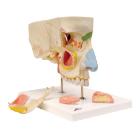
Model Nose With Paranasal Sinuses, illustrates the structure of the nose with the paranasal sinuses in the upper right half of a face in 1.5-fold enlargement, outside of the nose, with paranasal sinuses, differentiated by color, Dimensions : 26 x 19 x 24 cm
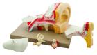
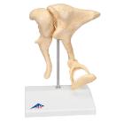
Model Ossicles 20X Bonelike, enlargement of original ossicles, created using micro CT, three smallest bones, joined to each other in the human body are located in the middle ear: malleus (hammer), incus (anvil) und stapes (stirrup), Dimensions : 17 x 12 x 21 cm
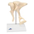
Model Ossicles Magnified 20 Times, joined to each other in the human body are located in the middle ear and are referred to as the auditory ossicles: malleus (hammer), incus (anvil) und stapes (stirrup), Weight: 0.385 kg, Dimensions : 17 x 12 x 21 cm